Dr. Trigo, José Manuel
Direktor für Onkologie, Forschung und Innovation

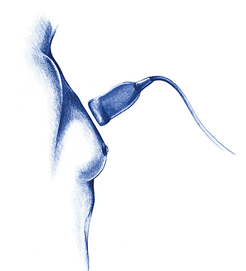
A non-invasive technique which uses ultrasound to obtain images of breast tissue. It is used as a complement to mammography and clinical examination.
Duration: 15 -30 min

When is it indicated?

Benefits

Limitations

Prepare for the test
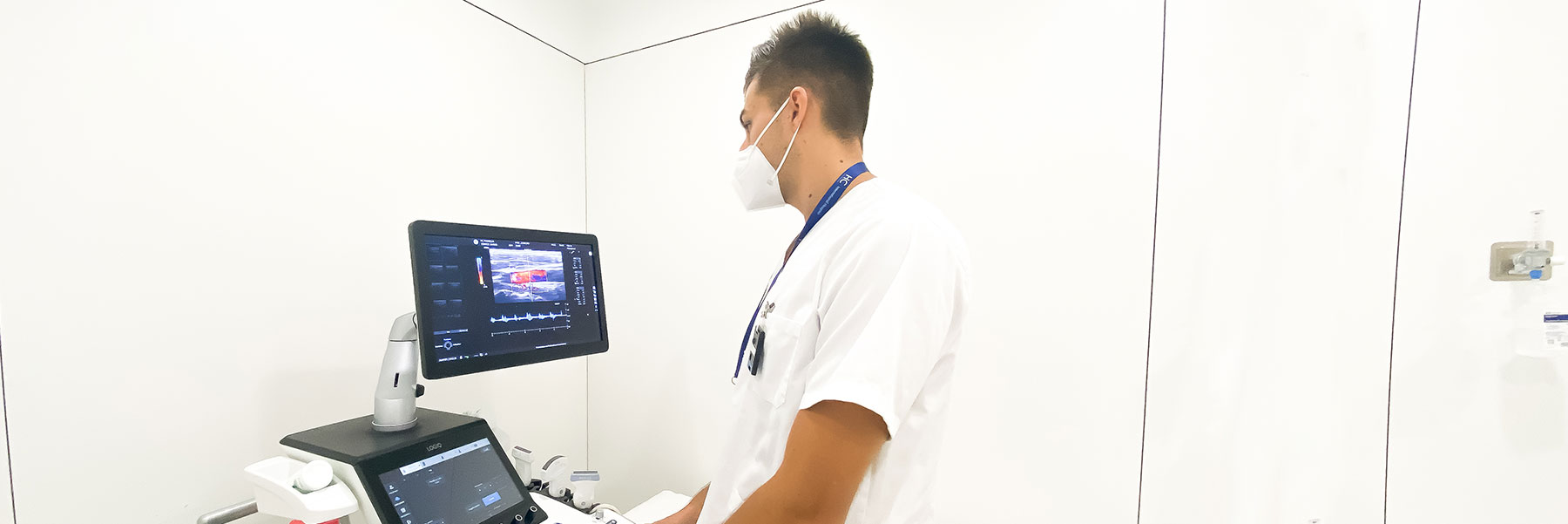
Breast ultrasound is a non-invasive imaging technique which uses sound waves to obtain detailed images of breast tissue. It is complementary to mammography, especially for the diagnosis of breast conditions. Although it is not a substitute for mammography, it is very useful when combined with clinical examination for the diagnosis and management of malignant and benign conditions.
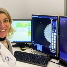
In our hospital, we use high-resolution ultrasound and high-sensitivity colour Doppler for more accurate results.
Breast ultrasound is primarily used to differentiate between solid masses and fluid-filled cysts which have been detected on mammography or during clinical examination.
It is also a key technique used to guide interventional procedures, such as cyst aspiration, FNA (fine needle aspiration) and targeted biopsy.
Although it is very useful in many situations, it is important to understand that ultrasound cannot detect microcalcifications, an important sign of early breast cancer which is best identified with mammography.
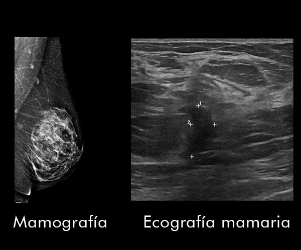
While mammography is the primary test to detect microcalcifications, a possible early indicator of breast cancer, breast ultrasound is most effective in assessing palpable masses or abnormalities not clearly seen on mammography.
Ultrasound is especially useful in women with dense breasts, young women, or those with breast prostheses, where mammography may not be sufficient. Together, both tests provide a more complete diagnosis.
The most common indications for breast ultrasound are:
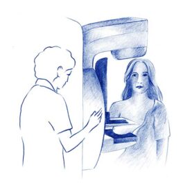
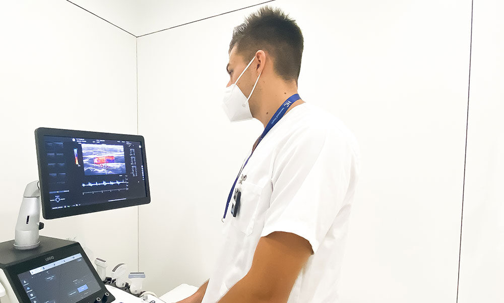
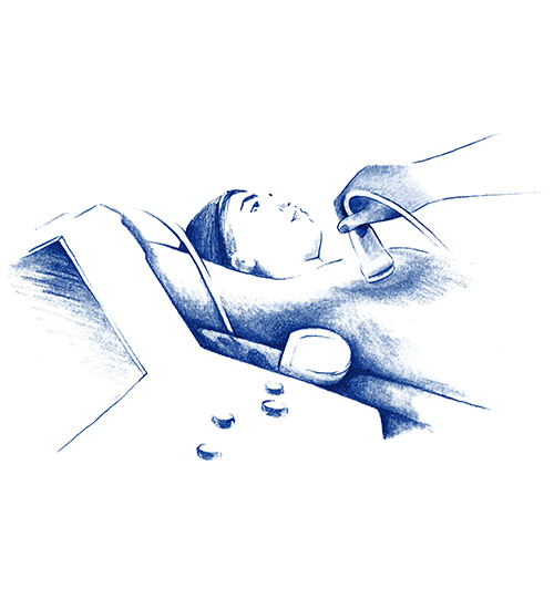
The procedure is simple and non-invasive. In our hospital, our state-of-the-art equipment includes high-resolution ultrasound with a special probe for detailed breast study. During the test, a gel is applied to the skin to improve contact with the probe, the probe moves over the breast to capture images in real time. The process lasts 15 to 30 minutes, it is completely painless.

Breast ultrasound is a valuable tool for assessing abnormalities detected by mammography or during clinical examination, this helps in the early diagnosis of breast cancer. However, it is not a suitable test for screening or mass detection of breast cancer, as it cannot identify microcalcifications, an early sign which appears in up to 50% of non-palpable cancers. Ultrasound is therefore used as a complement to mammography but should not replace it in the context of early detection.

Dr. Trigo, José Manuel
Direktor für Onkologie, Forschung und Innovation

Dr. Jiménez Rodríguez, Begoña
Facharzt für Medizinische Onkologie
Klinische Engagement bei Brust-und gynäkologischen Krebs

Dr. Sedano Ferreras, Paula
Spezialist für Radioonkologie

Dr. García Baltar, José Antonio
Especialista en Radiofísica Hospitalaria

Dr. Rebollo García, Natividad
HC Marbella Radiologie-Spezialist

Dr. Escobar, Ángela
Ginecología y Obstetricia, especialista en la Unidad de Mama

Dr. Bellinvia, Anna Alessandra
HC Marbella Radiologie-Spezialist

Dr. Arrazola, Tomás
Especialista en Farmacia Hospitalaria
Especializado en terapia contra el cáncer, certificado por la Sociedad Americana de Farmacéuticos de Hospital
Dr. López Ibor, Javier
Mental Health
Clinical and Forensic Neuropsychology

Dr. Di Mauro, Pietro
Especialista de la Unidad de Cirugía Plástica y Reconstructiva
Tel.: +34 952 908 628
+34 609 148 799
952908898 Onkologie
951829978 Bildgebende Diagnostik
951829947 Gynäkologie
952908897 Fertilitäts-Zentrum
951829947 Physiotherapie